Double-Injected Grass or Leopard Frog Dissection Specimen for Biology Labs | Vertebrate Anatomy
Due to a national shortage in the frog supply, these are currently unavailable.
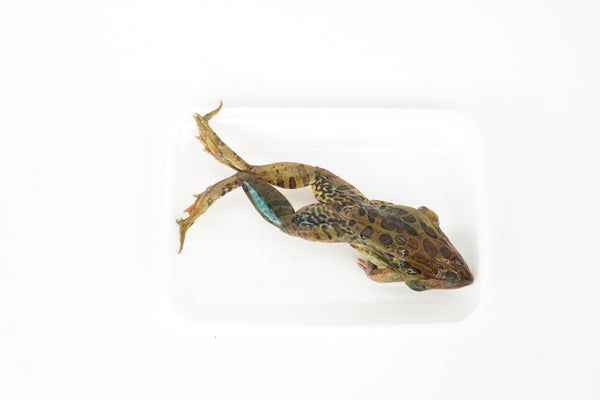
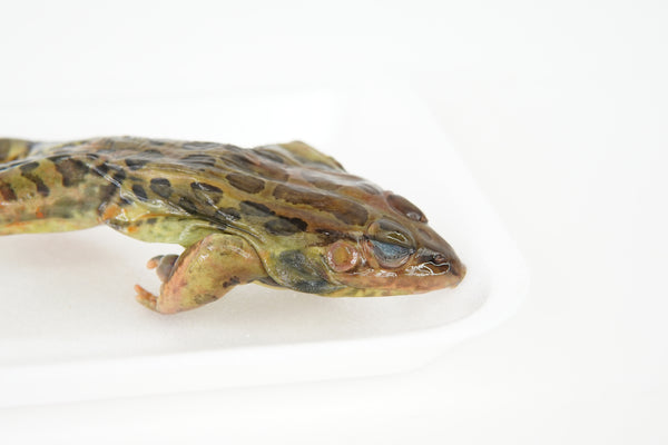
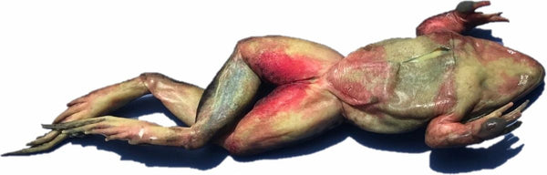
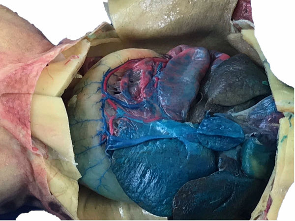
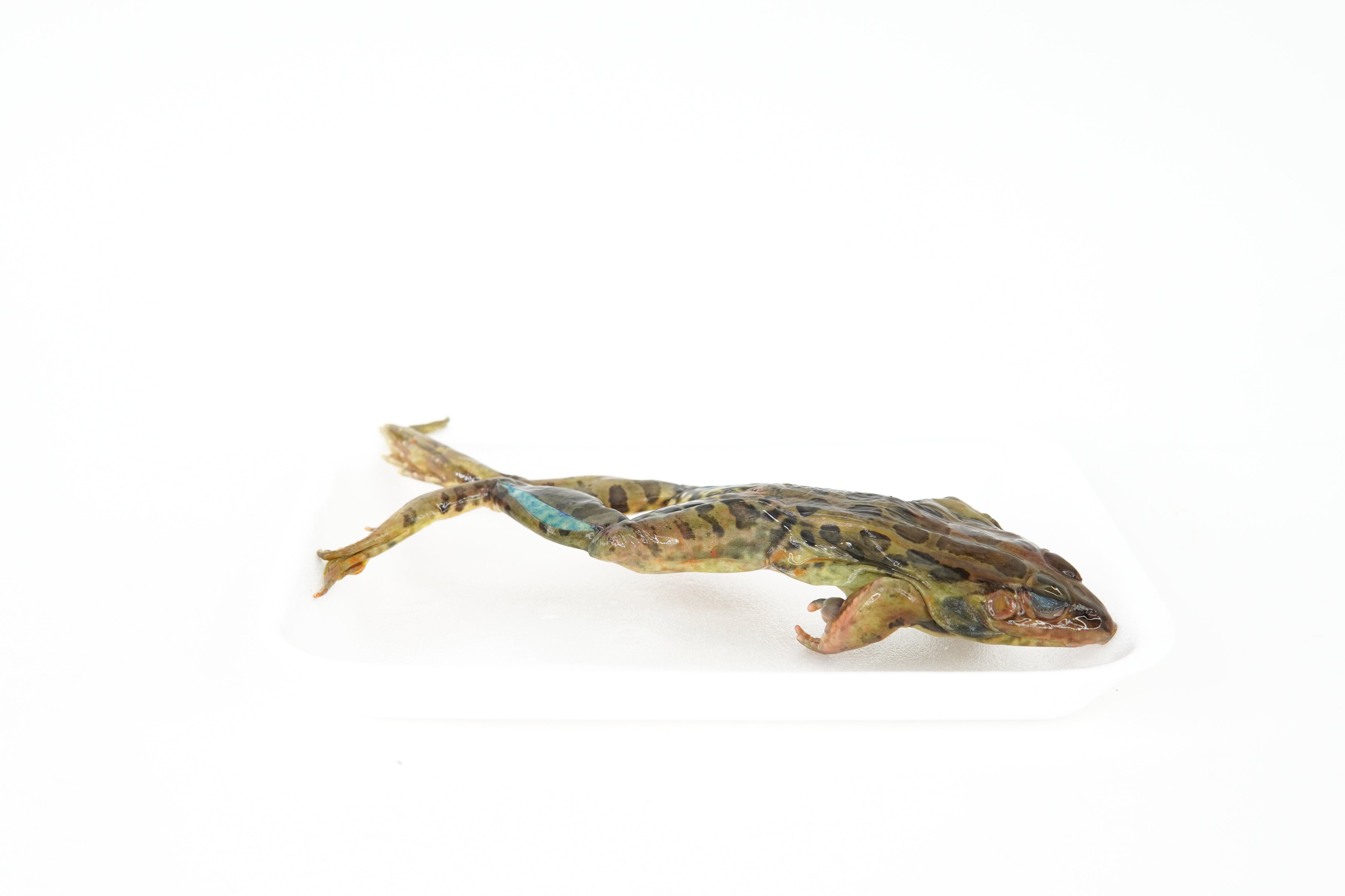
This double-injected grass or leopard frog dissection specimen is an excellent choice for activities requiring the dissection of a vertebrate animal and is widely used in middle school and high school biology labs to study vertebrate anatomy and organ system organization. Frogs are ideal model organisms because their body systems are clearly defined and easy to compare to other vertebrates. Each specimen has a body length of approximately 3–4 inches (legs not included) and is specially prepared with red latex injected into the arteries and blue latex injected into the veins to clearly display the circulatory system, allowing students to observe organs and blood vessels simultaneously during hands-on classroom dissection.
This preserved frog specimen for dissection supports instruction in comparative anatomy and physiology, making it a valuable specimen for anatomy labs, life science courses, and homeschool science programs. The double injection enhances visibility of the vascular system, helping students trace blood flow and better understand how circulation supports organ function.
Did You Know? Frogs do not chew their food. Instead, they use their eyes to help push food down their throat—when a frog closes its eyes while swallowing, the eyes press inward to assist the process.
Store specimens in an area of room temperature away from direct sunlight. Do not freeze or expose to high temperatures (more than 85ºF). Specimens will last up to 12 months when left unopened. After opening, the specimen should be placed in a zip-top bag and may be kept for up to one month.
Discounts available for purchases of 10 or more double-injected frog specimens!