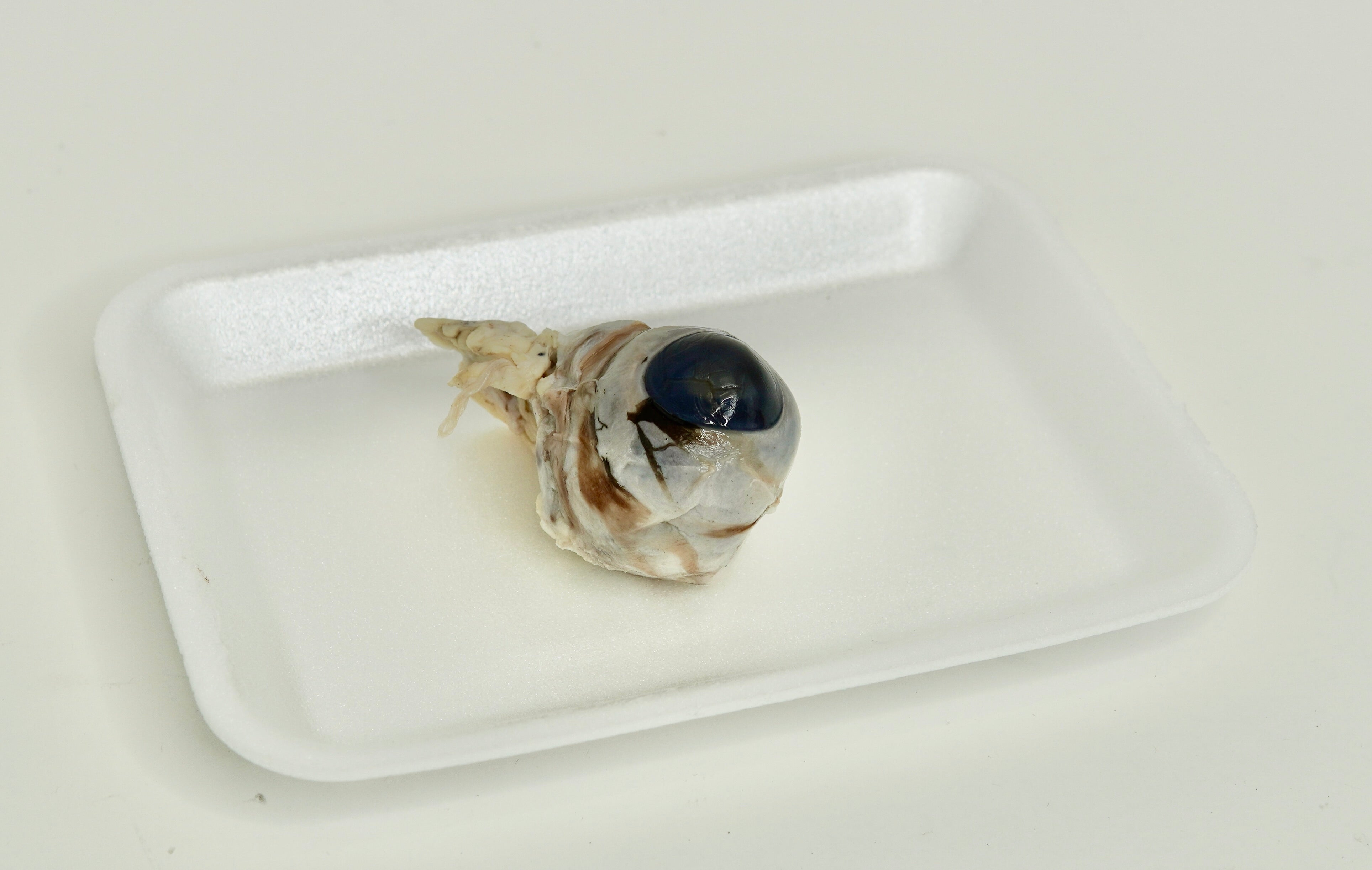
This preserved cow eye dissection specimen allows students to study the mammalian eye anatomy and understand how vision works in higher vertebrates. Commonly used in middle school, high school, and introductory college biology labs, the cow eye is an ideal model for exploring sensory organs because its size and structure closely resemble the human eye. Each specimen is preserved to maintain clear visibility of anatomical features, making it well-suited for hands-on classroom dissection focused on structure and function.
Cow Eye Anatomy Structures (External & Internal):
Sclera
Cornea
Iris
Pupil
Lens
Retina
Choroid
Optic nerve
Vitreous humor
Aqueous humor
Ciliary body
This preserved cow eye specimen for dissection supports instruction in vertebrate biology and sensory system function, making it an excellent choice for anatomy labs, life science courses, and homeschool science programs. Dissection specimens are initially preserved in formalin to fix tissues, then thoroughly rinsed and vacuum-packed in a low-odor holding fluid to ensure safe handling and long-lasting educational use. Each order includes one individually vacuum-packed specimen.
Did You Know? Cows have a reflective layer behind the retina called the tapetum lucidum, which helps them see better in low-light conditions and is the reason their eyes appear to glow when light shines on them at night.
Store specimens in an area of room temperature away from direct sunlight. Do not freeze or expose to high temperatures (more than 85ºF). Specimens will last 12 months when left unopened. After opening, the specimen should be placed in a zip-top bag and may be kept for up to one month.